
Can Surgery Treat Head and Neck Lymphedema? What LVA Offers Beyond MLD and Compression
Oct 05, 2026If you've gone through treatment for head and neck cancer, you already know the fight doesn't end when the tumor is gone. For a large number of survivors, the next chapter involves swelling, tightness, and heaviness in the face and neck that lingers for months or years, a condition known as head and neck lymphedema (HNL). This post breaks down what's driving that swelling, why standard therapy sometimes falls short, and how a microsurgical technique called lymphovenous anastomosis (LVA) is opening up a new treatment path.
What Is Head and Neck Lymphedema, and Why Does It Happen?
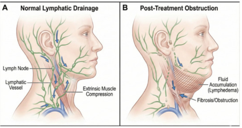
Head and neck lymphedema is one of the most common long-term consequences of head and neck cancer (HNC) treatment. Both of the pillars of HNC therapy, surgery and radiation, damage the lymphatic network. Surgical dissection severs lymphatic channels directly, while radiation triggers progressive fibrosis that stiffens and obstructs the vessels that remain. The result is the same either way: lymphatic fluid that can no longer drain efficiently pools in the surrounding soft tissue.
 (Harris et al., 2026)
Patients typically notice it first as a sensation of fullness or tightness rather than visible swelling. As fluid continues to accumulate, it becomes outwardly apparent puffiness in the submental region, jawline, or neck. When the swelling occurs internally rather than (or in addition to) externally, it can affect speech clarity and swallowing safety, since edema in the pharyngeal and laryngeal structures interferes with the coordinated movements those functions depend on.
- Periorbital involvement: swelling around the eyelids (palpebral lymphedema) can cause partial or complete ptosis, and in rare cases restricts eyelid closure enough to reduce visual acuity.
- Auricular involvement: edema affecting the external auditory canal can produce secondary hearing loss.
Both of these are easy to miss on a routine visual exam if the clinician isn't specifically screening for them, and both carry a real quality-of-life cost that goes beyond cosmetic concern.
The Current Standard: Complete Decongestive Therapy
According to the American Cancer Society's head and neck cancer survivorship guidelines, manual lymphatic drainage (MLD) is the recommended first-line intervention. In practice, MLD is rarely delivered alone, it's usually bundled into complete decongestive therapy (CDT), which combines:
- Manual lymphatic drainage
- Custom-fitted compression garments
- Therapeutic exercise
- Skin and wound care
CDT works, and it remains the foundation of conservative HNL management. But it asks a lot of patients. Sessions are frequent, often ongoing indefinitely, and compression garments for the head and neck region are notoriously difficult to tolerate and comply with long-term. For patients already managing fatigue, dysphagia, and the financial burden of cancer care, that ongoing time and cost commitment is a real barrier, which is exactly why interest in a more durable, less labor-intensive option has grown.
What Is Lymphovenous Anastomosis (LVA)?
Lymphovenous anastomosis is a supermicrosurgical procedure that gives lymphatic fluid a new way out. Rather than relying on the body's damaged lymphatic pathways, surgeons create a direct connection (a bypass) between a lymphatic vessel and a small nearby vein (venule), allowing lymph to drain straight into the venous bloodstream.
A quick clarification that's worth including in any patient-facing material: the "shunt" created in LVA is not a mechanical device, tube, or implant. It's a biological connection built entirely from the patient's own tissue, two microscopic vessels sutured together.
LVA has a well-established evidence base in limb lymphedema, where it's associated with measurable volume reduction, fewer infections (notably cellulitis), reduced long-term dependence on compression garments, and meaningful gains in quality of life. Its application to the head and neck is newer, but the underlying physiology and the surgical logic carries over directly.
How the Procedure Works?
The surgical procedure for lymphovenous anastomosis (LVA) begins with pre-operative mapping using ICG lymphography or high-frequency ultrasound to trace functional lymphatic channels and adjacent venules. Under general anesthesia, small incisions are made in natural skin creases or preauricular folds, followed by microscopic exploration using a high-powered operating microscope and superfine micro-instruments to isolate the target vessels. It is important to note that the term "shunt" in LVA does not refer to an artificial tube or implant; instead, it is a natural surgical bypass created by directly connecting two microscopic structures. The surgeon then selects an appropriate configuration and utilizes ultra-thin sutures (11-0 or 12-0 nylon) to stitch the vessel walls together. Finally, patency is verified to ensure proper fluid drainage into the venous system before the skin is meticulously closed with cosmetic sutures.
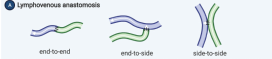
An operator of LVA should select an appropriate anastomosis type according to vessels available in a surgical field. There are 4 basic anastomosis types of LVA; end-to-end (EeE), end-to-side (EeS), side-to-end (SeE), and side-to-side (SeS).
Lymphatic supermicrosurgeon take 3 points into consideration; venous backflow rate, technical difficulty, and bypass efficacy. venous backflow rate is high in EeS anastomosis compared with EeE or SeE anastomosis, EeE anastomosis is technically easiest, and SeE and SeS anastomosis can bypass lymph fluid more efficiently compared with EeE and EeS anastomosis
(Gupta et al., 2021)
Why Timing Matters: Early and Immediate LVA
Lymphatic vessels don't stay operable forever. In early-stage lymphedema, the ducts are still relatively pliable; as the condition progresses, they become increasingly sclerotic (hardened and fibrotic), which makes successful anastomosis technically harder and less durable. This is the core argument for earlier surgical intervention rather than waiting until conservative therapy has clearly failed.
That logic has led to growing interest in immediate LVA, performing the bypass at the same time as the lymphadenectomy or dissection that puts the patient at risk for lymphedema in the first place, rather than waiting for swelling to develop and then treating it reactively. The data on immediate LVA is compelling: patients who receive it show meaningfully lower rates of lymphedema-related symptoms compared to patients who don't, evidence cited in the literature at a 26.53% relative risk reduction.
This prophylactic model (sometimes called immediate lymphatic reconstruction (ILR)) has already been studied extensively in breast cancer surgery, where it's reshaping how axillary dissections are approached. Head and neck cancer hasn't caught up yet. Current HNL management is still almost entirely reactive: therapy begins after lymphedema has already manifested, not before. That gap represents one of the more promising directions for future research and clinical protocol development in this space.
Is LVA Right for a Head and Neck Cancer Patient? Candidacy Considerations
LVA in the head and neck region isn't offered indiscriminately. It's generally considered when:
- Symptoms have progressed and conservative therapy (CDT) hasn't provided adequate relief
- The patient is younger and experiencing significant quality-of-life impairment
- There has been no cancer recurrence following completion of cancer therapy
That disease-free window isn't arbitrary, it reflects a genuine oncologic safety concern specific to this anatomical region. Lymphatic capillaries in the head and neck have fewer valves than those in the limbs, which means lymph flows more freely and with less directional control. Creating a new drainage pathway (the surgical "shunt") in a region with this kind of unrestricted flow raises a theoretical concern about facilitating cancer cell spread if any malignancy is still present. This is precisely why surgeons require confirmed, sustained remission before considering LVA. The follow-up period exists specifically to rule out local recurrence and nodal metastasis before creating a new route for lymphatic and, potentially, cellular traffic.
What Does the Evidence Show?
Outcome data for LVA in head and neck lymphedema is still an emerging body of literature, but the early results are encouraging:
- Facial contouring: the end-to-side technique achieved an average facial composite circumference reduction of 3.7%, with 75% of patients reporting a subjective improvement in skin tissue fibrosis.
- Symptom burden (external lymphedema): using the LSIDS-H&N (Lymphedema Symptom Intensity and Distress Survey — Head and Neck), scores improved from 1.11 ± 0.54 to 0.44 ± 0.66 over an average follow-up of roughly 15 months (P = 0.02).
- Objective staging (external lymphedema): using the MD Anderson Cancer Center Head and Neck Lymphedema (MDACC HNL) rating scale, patients improved from stage 2 down to stage 0 or 1a over a similar follow-up window (P = 0.008).
- Internal lymphedema and swallowing function: Swallowing Quality of Life scores improved from 130.5 ± 9.2 to 151 ± 19.8 over an average follow-up of roughly 21 months (P = 0.5)
LVA can be performed at different levels of the neck depending on where the lymphatic obstruction is located, which means (unlike compression garments and external MLD) it has the potential to address internal lymphatic dysfunction directly. That's a meaningful advantage, since internal HNL (affecting swallowing and airway-adjacent structures) is exactly the presentation that conservative therapy struggles to reach.
LVA is not a replacement for CDT, and it's not accessible to every patient or every practice.
- It is highly technically demanding, requiring surgeons with substantial microvascular reconstruction training and experience, this limits how widely available the procedure currently is.
- Therapeutic benefit is not immediate. Most protocols call for 8–12 months of follow-up before meaningful changes in the postoperative course become apparent, so patients need to be counseled on realistic expectations going in.
- Long-term outcome data, especially for internal/functional presentations, is still maturing.
That said, LVA remains a genuinely low-invasive option, and one that's proven effective for lymphedema across multiple anatomical regions, which is part of why interest in extending it to head and neck cancer survivors continues to grow.
Frequently Asked Questions
What is the difference between lymphovenous anastomosis and lymph node transfer? LVA connects existing lymphatic vessels directly to nearby veins to reroute fluid drainage. It does not involve transplanting lymph node tissue, which is a separate microsurgical technique (vascularized lymph node transfer).
How long does it take to see results after LVA? Most protocols recommend waiting 8 to 12 months of follow-up before evaluating the full therapeutic effect, since improvement tends to be gradual rather than immediate.
Can LVA be used for internal swelling that affects swallowing, not just visible facial swelling? This is still unknown. Depending on where in the neck the surgery is performed, LVA believes it address internal lymphatic dysfunction.
Does LVA replace the need for manual lymphatic drainage or compression garments? Not necessarily. LVA is typically considered after conservative therapy hasn't achieved adequate control, and many patients continue some form of CDT afterward, often at reduced intensity.
References
1- Mihara, M., Uchida, G., Hara, H., Hayashi, Y., Moriguchi, H., Narushima, M., Iida, T., Yamamoto, T., & Koshima, I. (2011). Lymphaticovenous anastomosis for facial lymphoedema after multiple courses of therapy for head-and-neck cancer. Journal of Plastic, Reconstructive & Aesthetic Surgery, 64(9), 1221–1225. https://doi.org/10.1016/j.bjps.2011.01.006
2-Lin, Y. S., Liu, C. J., & Chou, C. H. (2024). Lymphovenous Anastomosis for the External and Internal Types of Head and Neck Lymphedema: A Case Series and Preliminary Clinical Results. Plastic and reconstructive surgery. Global open, 12(6), e5872. https://doi.org/10.1097/GOX.0000000000005872
3- Yamamoto, T. (2013). Selection of anastomosis type for lymphaticovenular anastomosis. Journal of Plastic, Reconstructive & Aesthetic Surgery, 66(2), 207–208. https://doi.org/10.1016/j.bjps.2012.11.010
4- Hinson, C., Sink, M., Henn, D., Sammer, D., Zhang, A.Y., Billig, J.I., Chang, E. and Odobescu, A. (2025), Preventing Secondary Lymphedema: A Systematic Review and Meta-Analysis on the Efficacy of Immediate Lymphovenous Anastomosis. Journal of Surgical Oncology, 132: 717-726. https://doi.org/10.1002/jso.70046
5- Gupta, N., Verhey, E. M., Torres-Guzman, R. A., Avila, F. R., Jorge Forte, A., Rebecca, A. M., & Teven, C. M. (2021). Outcomes of Lymphovenous Anastomosis for Upper Extremity Lymphedema: A Systematic Review. Plastic and reconstructive surgery. Global open, 9(8), e3770. https://doi.org/10.1097/GOX.0000000000003770
6-Harris, M. K., Smith, J. D., Kim, J., Cai, W., Contrera, K. J., Chinn, S. B., Nilsen, M. L., Sridharan, S. S., & Spector, M. E. (2026). A Narrative Review of Lymphedema Following Head and Neck Cancer Treatment. Lymphatics, 4(2), 30. https://doi.org/10.3390/lymphatics4020030
Stay connected with news and updates!
Join our mailing list to receive the latest news and updates from our team.
Don't worry, your information will not be shared.
We hate SPAM. We will never sell your information, for any reason.